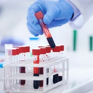
Book Your Test
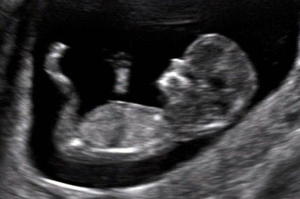
This test can be done when your pregnancy is between 11-13.5 weeks gestation (with the optimal time around 13 weeks) and it involves assessing the baby with ultrasound and taking measurements. These are entered into software developed by the Fetal Medicine Foundation along with the mother’s age and the results of the associated blood test. This program then allows us to calculate an individual risk of chromosomal abnormalities for each pregnancy.
This test is a risk assessment. It does not tell us which pregnancies are affected by chromosomal abnormalities. Approximately 5% of pregnancies will return as high risk and most of these will be normal, just as low risk does not entirely exclude an abnormality.
This screening method will detect approximately 88-93% of affected pregnancies.
Ultrasound is the term used for high-frequency soundwaves. Ultrasound examinations use these sound waves to produce a picture or image onto a screen showing the inside of your body.
An ultrasound is carried out by a Radiologist using a smooth, hand-held device called a transducer that they move across the body with a sliding and rotating action. The transducer transmits the high-frequency sound waves into your body. Different sound waves are reflected from different soft tissue, structures or parts in the body in different ways. These sound waves are converted to electrical impulses that produce a moving image displayed on a screen.
An ultrasound has many advantages. It is painless and does not involve radiation, which means it is very safe. The high-frequency sound waves ensure images show very high detail, capable of looking at the very tiniest parts of the body. Ultrasound can be carried out while there is movement, so it is excellent for the imaging of babies and children.
There are many reasons an ultrasound may be requested. For example, it is used to examine organs in the abdomen and other parts of the body. Colour Doppler ultrasound can be used to watch blood flow in any of the arteries or veins throughout the various parts of your body. High-resolution ultrasound can be used to evaluate the musculoskeletal system (muscle-, bone- and joint-related). Breast ultrasound is an important part of the assessment of any breast lump. Ultrasound can take high-quality images of most parts of your body, which makes it an excellent diagnostic test.
This will depend on the type of ultrasound that is requested. Listed below are some of the common examinations, with the preparation generally required.
All ultrasound examinations: Wear clothing that will provide easy access to the area that is being imaged. Bring any previous radiology examinations you have had with you (including ultrasound, magnetic resonance imaging, X-ray and computed tomography scans), so that they can be used for comparison and assessment.
Specific ultrasound examinations: Abdomen ultrasound: You will usually need to fast (have nothing to eat or drink) for 4-6 hours before the examination. This ensures there is no food or fluid covering the area that is to be examined. It also ensures the gall bladder is expanded to provide a clearer image.
Breast ultrasound: No preparation is required.
Female pelvis ultrasound: There are two ways to carry out this examination and the preparation will depend on which is used: a). Internal pelvic ultrasound – The best way to examine the pelvic organs in detail is to have a ‘closer’ look using a transvaginal (endovaginal) ultrasound where the ultrasound transducer is inserted into the vagina. b). External pelvic ultrasound – This is done in situations where an internal pelvic ultrasound is not appropriate, including children, or anyone who has not had an internal examination by their doctor. The ultrasound transducer is placed on the top of the lower abdomen (stomach area). To ensure that the inside of the pelvis area is seen clearly on the screen, a full bladder is required. You will need to drink 750 mL of water 1 hour before the procedure. Do not go to the toilet after drinking the fluid.
Thyroid ultrasound: No preparation is required.
Testes ultrasound: No preparation is required.
Musculoskeletal (muscle-, bone- and joint-related) ultrasound: No preparation is required.
Renal (kidney-related) ultrasound: You will need to drink 750 mL of water 1 hour before the ultrasound. Do not go to the toilet after drinking the fluid. Drinking the water will enlarge the bladder, enabling it and the surrounding internal areas to be examined.
Renal (kidney) arteries ultrasound: You will need to fast (have nothing to eat or drink) for 8 hours before the examination to ensure that the renal arteries are not covered by food or fluid.
Ultrasound is a safe examination that provides excellent imaging without any significant risk to the patient.
Ultrasound provides excellent imaging of the soft tissues of the body, and is a safe procedure that does not have the risks associated with imaging that uses radiation. There are no proven harmful effects of sound waves at the levels used in ultrasound when carried out in a proper clinical setting, such as a private radiology practice or hospital.
Rarely, a specific ultrasound contrast medium is injected into a vein of the arm to detect certain types of diseases or problems. If the radiologist feels that this will be useful, then this will be explained to you at the time of the examination. Ultrasound is mostly non-invasive, provides accurate imaging tests of the body, is readily available and is relatively inexpensive
An ECG is often used alongside other tests to help diagnose and monitor conditions affecting the heart.
It can be used to investigate symptoms of a possible heart problem, such as chest pain, palpitations (suddenly noticeable heartbeats), dizziness and shortness of breath.
An ECG can help detect:
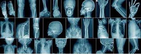
An X-ray study (also called a radiograph) is a type of medical imaging (radiology) that creates pictures of your bones and soft tissues, such as organs. X-rays use safe amounts of radiation to make these pictures.
We use digital computeted radiography technique at Plus 91 Diagnostics, where a digital cassette not film, transfers the image into an electric format. digital X-ray converts the X-rays that pass through the body more efficiently than film hence allowing less exposure for the same image quality. digital X-ray equipment provides superior image quality at lower radiation doses in comparison to older technologies.