Book Your Test

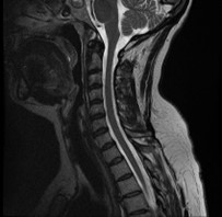
Evaluate symptoms such as pain, numbness, tingling or weakness in the arms, shoulders, or neck area. Find some types of chronic diseases of the nervous system. Diagnose tumors, bleeding, swelling, infection, or inflammatory conditions in the vertebrae or surrounding tissues.
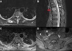
Diagnosis of back pain or other symptoms, such as numbness or tingling in the legs. Evaluation of any abnormalities or injuries to the spinal cord, nerves, vertebrae, and surrounding tissues such as vertebral fractures, slipped disc, etc.
A lumbar MRI can help detect and diagnose a variety of conditions affecting the lower spine, including problems with the spinal cord (such as infection or injury), degeneration of the discs and vertebrae (which can lead to nerve problems), and issues of the surrounding structures (like muscles and ligaments).

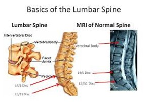
Joined at the very end of the sacrum are two to four tiny, partially fused vertebrae known as the coccyx or "tail bone".
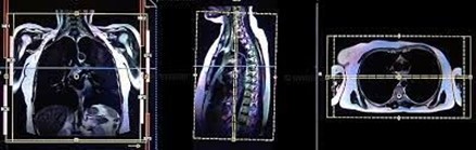
Magnetic Resonance Imaging (MRI) Screening of Whole Spine is used to visualise the spinal column (back bone) and the surrounding soft tissues like muscles using a small amount of radiation.
Ischemia/infarct,Vascular anomalies.Hemorrhage.Infection.Tumors and masses.Trauma and diffuse axonal injuries.Neurodegenerative disorders and dementias.Inflammatory conditions.
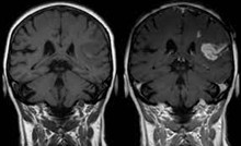
An MRI scan with contrast only occurs when your doctor orders and approves it. During the procedure, they'll inject the gadolinium-based dye into your arm intravenously. The contrast medium enhances the image quality and allows the radiologist more accuracy and confidence in their diagnosis.
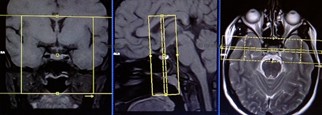
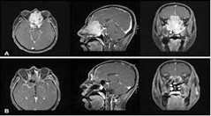
The most common endocrinological indication for performing a pituitary MRI was to evaluate for suspected prolactinoma, followed by evaluation of suspected Cushing's disease and hypogonadism.

Vertigo is a medical condition that results in the patient suffering from sudden bursts of excessive dizziness, which leaves them feeling off balance.
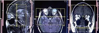
The commonest clinical indications for orbital imaging are proptosis/exophthalmos, diminished vision, enophthalmos, diplopia, leucocoria, pain, tumor and trauma. It is also used for evaluation in craniofacial developmental anomalies and epiphora.
Craniovertebral junction (CVJ) consists of the occipital bone that surrounds the foramen magnum, the atlas and the axis vertebrae.
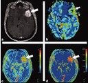
A brain perfusion scan is a type of brain test that shows the amount of blood taken up in certain areas of your brain. This can provide information on how your brain is functioning.
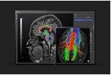
Diffusion-tensor imaging (DTI) is a noninvasive medical imaging tool used to investigate the structure of white matter. The signal contrast in DTI is generated by differences in the Brownian motion of the water molecules in brain tissue.
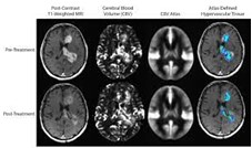
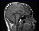
MRI protocol for brain tumor assessment is a group of MRI sequences put together to best approach CNS tumors in general. Note: This article is intended to outline some general principles of protocol design.
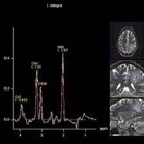
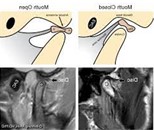
(Single Voxd1, MultiVoxd1)
Magnetic Resonance (MR) spectroscopy is a noninvasive diagnostic test for measuring biochemical changes in the brain, especially the presence of tumors. While magnetic resonance imaging (MRI) identifies the anatomical location of a tumor, MR spectroscopy compares the chemical composition of normal brain tissue with abnormal tumor tissue. This test can also be used to detect tissue changes in stroke and epilepsy.
MR is used to evaluate tumors and to assess for extension of an infectious process beyond the paranasal sinuses into the adjacent soft tissues. PET/CT is used for staging and restaging of head and neck tumors.
⇒ Provide an alternative to angiography, or avoid repeated exposure to radiation.
⇒ Clarify findings from earlier x-rays or CT scans.
⇒ Diagnose abnormal growths in the chest.
⇒ Evaluate blood flow.
⇒ Show lymph nodes and blood vessels.
⇒ Show the structures of the chest from many angles.

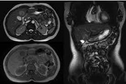
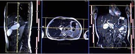
An MRI of the liver can show the structure of the liver, as well as atypical growths.
⇒ Blood flow in the abdomen.
⇒ Blood vessels in the abdomen.
⇒ The cause of abdominal pain or swelling.
⇒ The cause of abnormal blood test results, such as liver or kidney problems.
⇒ Lymph nodes in the abdominal.
⇒ Masses in the liver, kidneys, adrenals, pancreas, or spleen.
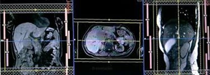
Identification of congenital anomalies of the cystic and hepatic ducts.post-surgical biliary anatomy and complications. ...pancreas divisum. anomalous pancreaticobiliary junction.
choledocholithiasis. ...biliary strictures. ...chronic pancreatitis. ...pancreatic cystic lesions.
In the third phase, the contrast is flushed out of the portal veins and helps to visualize the veins. This MRI examines the physiology of the liver to determine whether there are any liver abnormalities or the existence of any liver tumor (growth/cancer).
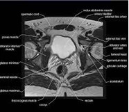
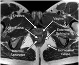
Abnormal vaginal bleeding.
A mass in the pelvis (felt during a pelvic exam or seen on another imaging test)
Fibroids.
A pelvic mass that occurs during pregnancy.
Endometriosis (usually only done after ultrasound)
Pain in the lower belly (abdominal) area.
MRI has very high contrast resolution, which means that it can see the different layers of the bowel wall and surrounding structures. It also is able to use different sequences to specifically look for any small areas of inflammation within the bowel or more commonly the deeper structures.
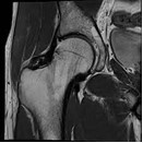
Typical indications include pain in the hip and/or buttock, hip-related groin pain, decreased range of motion, limping and comprise the following: osteonecrosis of the hip. transient bone marrow edema syndrome. femoral insufficiency or stress fracture.
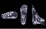
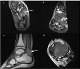
An MRI foot scan is a diagnostic tool used to image the structures of the foot and ankle. You might need an MRI foot scan if you experience any of the following symptoms: pain in one or both feet, swelling, redness or warmth, numbness or tingling, stiffness, or decreased sensation in the feet.
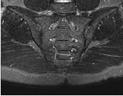
MRI is the most sensitive imaging technique to detect sacroiliitis. It is the only imaging modality that can reliably reveal bone marrow edema and inflammation around the sacroiliac joints and is comparable to low dose CT for demonstrating erosions and ankylosis
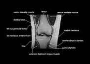
knee pain, weakness, swelling or bleeding in the tissues in and around the joint.
damaged cartilage, meniscus, ligaments or tendons.
sports-related knee injuries, such as sprains and torn ligaments, cartilage, or tendons.
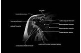
Tumors or infections.Bone fractures. Rotator cuff tears. Arthritis or degenerative joint diseases. Shoulder joint dislocation. Decreased in the motion range. Injuries due to sports.
Bone fractures.
Ankle sprain.
Midtarsal sprain.
Stress fractures.
Suspected chondral injury.
Ankle impingement.
Achilles tendinopathy.
Plantar fasciitis.
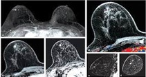
Breast MRI is indicated for the evaluation of unilateral metastatic axillary lymphadenopathy with an unknown primary malignancy. MRI used in these instances is able to identify a primary breast malignancy in 62–86% of patients, allowing targeted systemic therapies and often breast conservation.
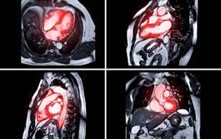
Coronary artery disease.
Cardiac masses and thrombi.
Pericardial abnormalities.
Cardiomyopathy.
Congenital cardiac disease.
Arrhythmia.
Aortic disease.

Magnetic resonance angiography (MRA) is a very valuable tool in the routine evaluation of patients with stroke syndrome. It provides powerful noninvasive imaging of the cervical and intracranial vessels allowing the detection and the diagnosis of vascular anomalies.
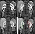
Nonenhancing hyperattenuating cysts that are larger than 3 cm or completely intrarenal are included in this category (1). According to one study, 95% of these lesions are benign and therefore have a low probability of being small renal cell carcinoma and a low chance of metastasis (6)
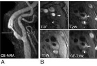
To assess the quality of images obtained from time-resolved MRA together with the accuracy of this technique in diagnosing vascular diseases and the usefulness of haemodynamic information provided by this method.

CT angiography is a type of medical test that combines a CT scan with an injection of a special dye to produce pictures of blood vessels and tissues in a part of your body.

What can an MRI of the prostate show? Not only is a prostate MRI as accurate as a biopsy at detecting prostate cancers, but it can also tell how advanced the cancer is and if it has spread to other parts of the body.

Magnetic resonance enterography is an imaging test that lets your doctor see detailed pictures of your small intestine.

cervical spine MRI also can help doctors: Evaluate symptoms such as pain, numbness, tingling or weakness in the arms, shoulders, or neck area.
Magnetic resonance imaging (MRI) is one of the best diagnostic tools for identification of TMJ pathology, allowing evaluation of TMJ disc position, morphology, mobility, extent of joint degenerative changes, inflammation, and presence of connective tissue/autoimmune diseases.

A heart MRI is a scan of your heart in which radio waves and magnets create images without anything you can see or feel going into your body.