Book Your Test

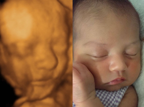
Optimum at 24-30 weeks
We can make this special experience even more exciting by providing the first 3D 4D glimpse of your new baby.
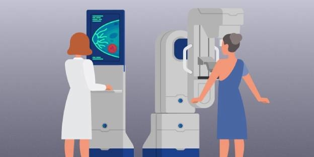
A mammogram is an x-ray procedure used specifically to X-ray the breasts. Every effort will be made by the Mammographer to ensure you are as comfortable as possible during the examination. Patients will be able to return to normal activities immediately after the procedure.
The mammography procedure is used by doctors and specialists to diagnose breast cancer, and various other diseases of the breast, for example, fibroid adenoma. An ultrasound examination may also be recommended to obtain further information. This is usually done when breasts are especially dense.
Mammography can detect tumors that are too small to be felt and have reduced the rate of terminal cases of breast cancer in patients aged between 40 and 70.
Patients who are considered at high risk of developing breast cancer (i.e. due to a family history of breast cancer) may also be advised by their doctor to undergo regular testing.
There is a small amount of radiation used; however, the benefits outweigh the risks from the radiation.
Your details will be checked, and your examination carried out by a radiographer who has specialized in breastwork or more commonly called a Mammographer. You will be asked to undress from the waist up. It is recommended that you wear a loose, comfortable, two-part outfit. The whole appointment takes less than half an hour and the actual mammogram will take around 5- 10 minutes.
This scan is performed in early pregnancy to determine the age of the pregnancy (also known as the gestational age) using measurements. The measurements are from one end of the fetus to the other, known as the crown-rump length, or CRL.
This scan is usually done if you are unsure about the dates of your last menstrual period (LMP) or have irregular periods. If you have been breast feeding or have recently stopped the oral contraceptive pill you may have had an unusual cycle so you may not be sure of your LMP.
This scan may also be performed also if you have an early pregnancy and have bleeding or pain (even if you have already had a scan or are sure of your last menstrual period) to assess if the pregnancy progressing normally.
A scan performed between 7-11 weeks pregnancy/gestation is accurate to within 3 days. What else does it show?
Most of the images can be obtained by transabdominal ultrasound (this involves scanning through your lower abdomen) using a probe and gel on your skin. This requires a full bladder. Sometimes if the pregnancy is early or better images of your uterus or ovaries is required a transvaginal ultrasound scan is required. This involves an internal probe being gently placed into the vagina with a sterile protective sheath (usually less discomfort than a PAP smear) This allows better images as the probe lies closer to these structures.
This depends how far along your pregnancy is. Even if you have a positive pregnancy blood or urine test it may be too early to see signs of a pregnancy on ultrasound. Before 5.5 weeks a small fluid sac may be seen in the uterus without sign of a fetus or a fetal heart beat. In these situations a repeat scan one to two weeks later may be necessary to confirm a viable pregnancy and provide accurate dating. Your doctor will also usually correlate the results with blood tests (serum BHCG) By 6-7 weeks the fetus is usually seen clearly (often requires transvaginal scanning) and its heart beat can be visualised.
This ultrasound examination (also known as an anomaly scan) provides a detailed scan of your developing baby and is considered to be routine practice for all pregnant women in Australia. At Spectrum, we recommend booking your morphology scan after 19 weeks gestation.
At this stage the sonographer and doctor are able to look at the baby’s anatomy (this involves a detailed examination of the baby’s head/brain, face, heart, stomach, kidneys, bladder, spine and limbs) They can also check the position of the placenta in relation to your cervix, the length of the cervix and fluid around your baby.
This is thought to be the optimal time to visualise the baby’s anatomy, allowing accurate assessment of the internal organs and limbs. The timing of the scan also allows further testing should an abnormality be found.
Despite advanced technology and the very experienced medical professionals at our practice ultrasound does not detect all abnormalities a baby can have. There are some abnormalities that are only evident later in the pregnancy such as some cardiac defects and bowel abnormalities There are conditions such as cerebral palsy and chromosomal abnormalities that cannot be detected at all. This scan provides reassurance for you and your doctor however dos not guarantee a normal baby or that you will have no pregnancy complications.
Fluid in your bladder provides a “window” to get better images on your scan. It also helps push the uterus (womb) up from your pelvis and bowel out of the way to better visualise your baby, uterus and cervix. We suggest you empty your bladder an hour before your appointment and drink ¾ of a litre of water and then try not to empty your bladder until after your appointment (your bladder does not need to be so full that it is painful)